|
For kidney anatomy, it is important to keep in mind that the kidneys are retroperitoneal organs. This means they do not have a mesentery and are attached to the posterior body wall. Some other retroperitoneal organs include the aorta, esophagus, adrenal glands, and rectum.
The heart delivers blood to the kidneys via the renal artery. That blood contains oxygen and nutrients such as electrolytes, amino acids, and glucose. It also contains waste products, like urea, unused electrolytes, and byproducts of cellular respiration, to be excreted in the urine.
Blood from the renal artery travels through two capillary beds, the vasa recta and peritubular capillaries, to connect to the renal vein. The vasa recta delivers oxygen to the kidneys whereas the peritubular capillaries collect nutrients from filtration to be recycled. The renal vein collects the nutrients and oxygen the kidneys do not use to be returned to the body.
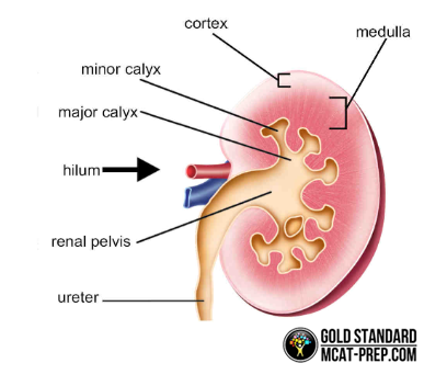
At the very exterior of the kidney is the renal capsule, a transparent membrane that encloses the kidney to maintain its shape and protect it from damage. Beneath the capsule is the renal cortex or shell-like portion of the kidney. The darker, middle portion inside the cortex is the renal medulla. And the medulla is divided into sections called renal pyramids which contain nephrons.
The nephron is the smallest functional unit of the kidney. Nephrons extend between the renal cortex and renal medulla to facilitate filtration as well as collection. Each kidney contains approximately one million nephrons. The renal calyces (singular: calyx) have minor and major portions for collecting urine from the renal pyramids. This is the first place where urine is found in the kidney.
Renal calyces collect into a central, funnel-shaped kidney area called the renal pelvis. From there, urine drains into the ureter, a tube through which urine travels to reach the bladder for excretion. Finally, the region of the smaller curvature of the kidney where the renal artery enters and the renal vein exits with the renal pelvis is referred to as the renal hilum.
Of course, the MCAT will also question your knowledge of renal physiology and its responses to changes in homeostasis. Nevertheless, understanding kidney anatomy will give you a firm foundation to build on for test day.
|